Monday, January 30, 2017

pGLO Lab

        In the pGLO lab, we added plasmids to E. coli bacteria containing araC, GFP, and ampicillin resistance genes. After heat shocking the bacteria, we plated them onto luria broth with ampicillin nd arabinose and waited for the colonies to grow.
        Though my group's bacteria failed to receive the plasmid successfully due to possible errors in adding the plasmid and heat shocking, we recorded data from a more successful group to analyze.
Our failed plates (note no GFP in bacteria seen under black light)

Another group's successful plates
(from left to right: -pGLO with LB,
+pGLO with LB and amp,
+pGLO with LB, amp, and arabinose)

Plate
Number of Colonies
Color of colonies under room light
Color of colonies under UV light
- pGLO LB
1 large colony with incoherent boundaries
Murky gray
Yellowish gray
+ pGLO LB/amp
9 large colonies
Murky gray
Yellowish gray
+ pGLO LB/amp/ara
7 colonies
Murky gray
Bright green
        There were likely about 10,000,000 bacteria in each 100 uL E. coli sample plated, assuming about 100,000,000 in each milliliter. This estimate takes into account the millions of bacteria that exist everywhere and gives a reasonable number without a reference, though an equally large number is possible as well.
        Out of these, a very small number of bacteria (i.e. around 1,000,000) successfully received the plasmid. These transformed bacteria had the pGLO gene as well as ampicillin resistance, allowing them to be isolated in the luria broth, as the ampicillin killed off the rest of the bacteria. In addition, arabinose acted like a trigger to activate the GFP intron in the pGLO plasmid, allowing the bacteria to produce GFP when the sugar was present. Where arabinose was absent, (as in the +pGLO LB/amp plate) the gene was not expressed; in the plate with the trigger sugar, the gene was expressed, and the bacteria glowed.
        This kind of genetic engineering is very useful and can be applied in a variety of places. GFP is used in many areas, including as a cell marker, in which it is added to plasmids of interest to gauge how many bacteria have successfully taken in the plasmid; as a transcription reporter, in order to monitor the expression under a certain promoter; and in FACS (fluorescence-activated cell sorting), to separate different cells based on their fluorescent signals. Furthermore, genetic engineering involving the enzyme Cas9 in CRISPR editing can be done in order to modify the genome of different organisms, including humans, with low costs and equipment. This can been done to change the inheritance of traits or characteristics of embryos, and is currently being studied.

Thursday, January 19, 2017

Gel Electrophoresis Lab

        Though there are natural alternatives, artificial food colors can be preferable to both consumers and manufacturers. They are much easier to mass-produce and obtain, and also include a wider range of bright colors (or colors that make food seem fresher) that can seem more appealing to consumers. Though I usually tend to avoid foods and snacks with dyes, I have seen sodas that contain different dyes, as well as chips (e.g. Doritos) that contain dyes like Yellow 5 and Yellow 6. Additionally, one large source of artificially dyed foods is dog food: manufacturers tend to add artificial food colors like Yellow 5, Blue 2, Red 40, and Yellow 6. This is likely to create a more “nutritious” appearance that may sway consumers.
        In our DNA electrophoresis lab, we ran dyes found in various common candies. We used the Red 40, Yellow 5, Yellow 6, and Blue 1 reference dyes to compare our dyes to. Observing the molecular structures of other dyes, we can see that they have similar structures to the four reference dyes we used, with analogous substituents (e.g. phenyl, hydroxyl, carbonyl); these similar structures would lead to them to migrate similarly in gel electrophoresis. For example, carminic acid is similar to Yellow 6, betanin acid bears resemblance to Yellow 5, fast green FCF is like Blue 1, and citrus red 2 resembles Red 40.
        Our dyes ran mostly as expected, with the orange dye being Yellow 6, and the other dyes turning out to be their corresponding reference dyes. However, our green dye separated into 2 different bands when we ran the gel electrophoresis. This green was actually a mixture of a blue and a red dye (Blue 1 and Red 40), since green dyes are likely harder to produce.
Final gel results (4 test dyes on left, 4 reference dyes on right)
        The colored dye solutions we used also migrated different distances, and this effect was contributed to by multiple factors. First of all, the larger molecules are less flexible and will be hindered more by the gel’s channels. Also, the molecule concentrations would make a difference: a dye with a higher concentration might advance slower than another dye with a lower concentration.
Gel electrophoresis setup
        The movement of the dyes through the gel is facilitated by their negative charge and the voltage of the electricity. Their charge makes them attracted toward the anode, and smaller molecules can advance further, because they are less obstructed by imperfections and twists in the gel. DNA molecules, which are commonly used in gel electrophoresis, would also separate like the dyes we used. DNA of a lower dalton size would advance the furthest, while the largest molecules would advance much slower and thus traverse less distance.

Tuesday, January 10, 2017

New Year Goals

        This semester I will strive to improve upon last semester's performance in several specific ways. In order to attain more understanding in biology, I will study and investigate more into each unit. I will make connections to different aspects of biology and find in-depth information about each one, enhancing my understanding about the specific subject and its place in the world of biology.
        Additionally, I will improve my expository writing in English, which will entail forming stronger and better substantiated points and theses, as well as creating a conclusion that better wraps up the essay. This will allow me to better express my opinion and evidence in future letters or pieces of writing.
        In order to gauge my progress, I will reflect (perhaps sometimes physically) on my accomplishments toward my goal in each unit or subject: in Biology, this would involve connecting the topic to other concepts, while in English I would keep track of each facet I expressed successfully or included after each essay or other practice opportunity.

Thursday, December 15, 2016

Unit 5 Reflection

        In this unit, I learned about processes involved with the essential nucleic acids: DNA and RNA. I learned about how DNA has an antiparallel, double helix structure made of nucleotides. These nucleotides have nitrogenous bases, which include the purines adenine and guanine, and the pyrimidines cytosine and thymine.
Structure of DNA

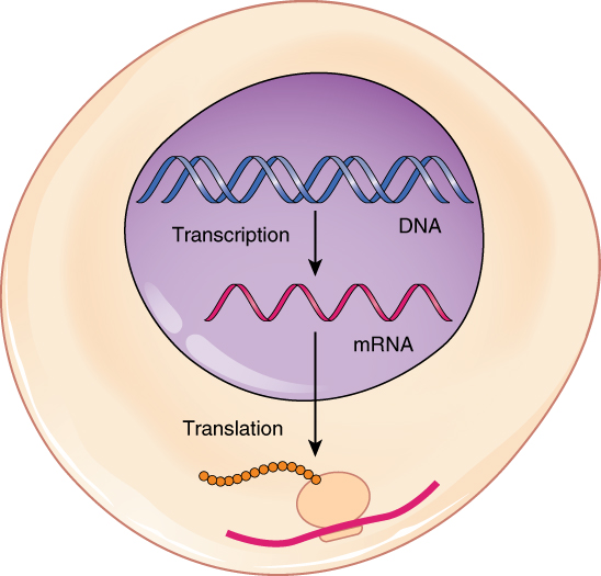
They pair to make a code, which can be replicated through semi-conservative replication during interphase, or be transcribed by helicase and RNA polymerase to create mRNA used in protein synthesis. The mRNA is translated into amino acids in a ribosome, and the resulting protein is sent to use in the cell or body. However, mutations can cause significant changes in DNA that lead to radically different proteins being produce, and this can interfere with processes. Additionally, gene expression is regulated by transcription factors and repressors, keeping certain genes from being read by RNA polymerase. This controls the production of certain proteins, such as lactose, from being produced unless they are needed. In general, much of this was a review for me, but there were also times where I took longer to understand new concepts that were presented, such as how operators work in gene expression and regulation. On the other hand, the structure of DNA and protein synthesis were not too challenging to grasp, especially since I had encountered them before in science classes, though the practice was very helpful.
DNA extracted during the DNA extraction lab

        Throughout the unit, I found that I learn better when asking questions and clarifying more complex parts of the content. I discovered that for topics like gene regulation and expression, I tended to understand the material and process much better after the in-class discussion the next day. As a student, I would consider myself having improved, as I learned how to garner more information and skills from what is presented; in essence, I am not only smarter now than a month (or the duration of the unit) ago, but I have improved at improving. (Δ(Δk)?) I think the strategy of physically typing and creating a study guide is an effective one: through writing down the material, one is reviewing and remembering perhaps nuances that were forgotten over the course of a few months. Next semester, I will try to utilize this method to synthesize material better and continue to learn more. (and learn more about learning)

Tuesday, December 13, 2016

Protein Synthesis Lab

        In this lab, we modeled the processes involved in protein synthesis. To make a protein, on goes through transcription and translation. First of all, we copied down the DNA strand and transcribed it into mRNA, replacing thymine with uracil. This is what RNA polymerase does in the nucleus. Then, we translated the mRNA by splitting it into codons and finding the corresponding amino acid for each codon. This process, representing the tRNA's work in the ribosome, produced the primary structure of the protein.
Diagram of protein synthesis
https://commons.wikimedia.org/wiki/File:0328_Transcription-translation_Summary.jpg

        The mutations that seemed to have the greatest effect were the frameshift mutations, insertion and deletion, while the mutation that seemed to have the least effect was substitution. For the frameshift mutations, the location of the mutation was also very influential: a mutation near the beginning of the sequence would alter (shift over) almost every codon, while one at the end would change very little. This is due to frameshift mutations affecting every codon after it.

Some possible mutations in DNA
https://en.wikipedia.org/wiki/File:Chromosomes_mutations-en.svg

        I chose the frameshift mutation as what my data indicated would cause the most damage to the gene. This mutation, unlike other mutations we tested, had a chain reaction effect that rippled across and changed the frames of every codon after it. This could lead to devastating consequences, such as a stop codon in the beginning or completely different amino acids. As mentioned earlier, this effect is especially predominant if the mutation is at the beginning, as it would change everything after it.
Effect of a frameshift mutation (insertion)
https://www.flickr.com/photos/yourgenome/26855221022

        Because of the direct connection between DNA and proteins, and the influence proteins have in the body, dangerous DNA mutations can wreak havoc and create abnormalities in daily processes such as blood flow, digestion, and respiration. For example, cystic fibrosis is caused by a mutation in the CFTR gene. These mutations prevent chloride ion channels from functioning properly, and unusually thick mucus is produced, which clogs airways.
Diagram of cystic fibrosis
https://www.flickr.com/photos/yourgenome/26855222462

Monday, December 5, 2016

Human DNA Extraction Lab

        In this lab, we asked how DNA can be separated from cheek cells to study. We found that significant amounts of DNA can be extracted as a visible precipitate. The thread-like structure could be seen suspended in the isopropanol alcohol, after the layer of nonpolar alcohol was added on top of the solution with DNA. It has been proven that through homogenization, lysis, and precipitation, one can extract DNA. This is done through breaking down the membranes and nuclear material with polar liquid, lysing the membranes, emulsifying the proteins and lipids, and breaking down the histones in the DNA. Then, by adding a polar substance as a layer, the DNA will fall out of solution at the interface and become a precipitate. Our data supports this claim and is the result of such a process, using salt as a precipation facilitator, detergent as a disruptor, and pineapple juice as a catabolic protease to help break down different structures in the cell and free the DNA.
DNA precipitates into the ispropanol alcohol
        Some errors people in our lab group made led to alcohol and solution mixing, thus precluding the DNA from becoming a precipitate. In one experiment, the inversion to mix detergent with the solution was done too quickly and violently, thus leading to bubbles forming and disrupting the liquid surface. This would have kept the DNA from successfully precipitating at the interface, and could have mixed up or damaged the DNA strands in the solution. Another error was not measuring very precisely, which lead to a variety of results, some unsuccessful. We simply estimated amounts (e.g. pinch of salt), which may have led to imprecise measurement and thus a failed extraction with a certain substance being too dilute or concentrated. In a future experiment, I would suggest making sure to mix slowly, which would keep the solution from forming many bubbles. Additionally, keeping measurements precise, such as using measuring spoons and graduated cylinders, would give less variation and a higher success rate in general.
Precipitation is unsuccessful due to violent inversion
        This lab was done to demonstrate how DNA can be extracted from inside the nucleus of a cell. Moreover, I also learned how the membranes, histones, and nuclear material must all be broken down before the DNA can be extracted. This helped me understand how much protection the DNA has, due to it holding the genetic information. Not only does it never leave the nucleus (mRNA copies and leaves instead), there are many layers of membranes to keep it from being damaged by substances outside the cell. Outside of these concepts, based on my experience from this lab, I also now know how one might break down cell membranes to access, not only the DNA, but any organelle one might be studying using the techniques of homogenization, lysis, and precipitation.

Wednesday, November 30, 2016

Coin Sex Lab and Unit 4 Reflection

        In the coin sex lab, we simulated the inheritance of traits on different genes through crosses. This effectively models genetic concepts, as the flipping of coins represents the random recombination of alleles, and thus creates plausible results that conform to the combinations found in a punnett square. We tested a cross with sex chromosomes, autosomal inheritance, x-linked inheritance, monohybrid crosses, and a dihybrid cross. Across the board, though our results and ratios did not always completely match the expected results, such a discrepancy can easily be explained by the small sample sizes (10-16 flips), and our results all fit within the possible genotypes from meiosis. Using probability to predict offsprings' traits is limited in that in the real world, as the results often vary from both randomness and environmental traits (here factors that affect the flip result).
Data for dihybrid cross
        As expected, the X-linked colorblindness cross yielded mostly affected males, and the dihybrid cross yielded different genotypes with more double heterozygous and few double homozygous. In the dihybrid cross, BbEe brown haired, brown eyed individuals were crossed, which is expected to produce 9:3:3:1 phenotypic ratio. Our results produced a 14:1:1:0 ratio, which is close to expected, and this high brown-brown expected value is due to there being more combinations that result in it. Dominant alleles dominate others, so the double dominant genotype would be much more common. This understanding can be directly related to life, as dominant traits are observed much more in the phenotypes than recessive ones, and this can be used to understand how recessive traits such as blue eyes or blond hair are passed down through generations even if masked. This can explain where people around you attained such unique characteristics as the ones they have.
        In this unit, we explored concepts like these, such as the cell cycle (interphase, mitosis, cytokinesis), the process of meiosis, how asexual and sexual reproduction each have their costs and benefits, the difference between haploid and diploid cells, Mendel's experiments with pea plants, the principle of inheritance, Mendel's Laws of Segregation and Independent Assortment, genotypes and phenotypes of traits, how dominant alleles mask recessive alleles, the usage of Punnett squares, incomplete dominance and codominance, how genes affect one another through epistasis, and the multiple genes that contribute to polygenic traits.
Diagram and punnett square of a dihybrid cross
        Some of this material was a bit challenging and confusing to understand, such as the complex probabilities of X-linked traits. However, after practicing and learning about how these alleles are passed along differently in males and females that guarantee certain things, I have grasped a better understanding of it. After lectures and completing the labs and infographic, I could make more connections between the different aspects of genetics, and understood better how Mendel's laws make the principles and complications possible. In addition, I would like to explore some of the more convoluted processes a bit more. For example, how do the spindle fibers in meiosis work? How are they produced and how do they extend and latch onto chromosomes to pull them apart? Also, in DNA replication, how exactly are the strands replicated? Are the leading and lagging strands copied differently due to their direction?